

Chaperonin Assembly and Substrate Folding
Oksana Sergeeva
2010-2014; PhD student
oksana.sergeeva@gmail.com
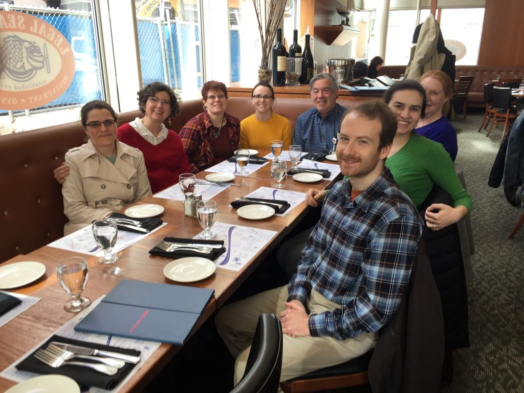
My years in Jon’s lab were some of the best years in my life. Not only did I love living in Cambridge and attending grad school, but Jon created a special environment in the lab that made it possible to be very productive in a more-or-less stress-free way. Unlike other labs, he didn’t pressure us to produce results, but to do good, thoughtful science. It wasn’t always the most glamourous science, but I am very proud of the work we did in Jon’s lab. My time in the early 2010s was during a downturn in the lab; Jon’s lab was winding down and decreasing in size. This caused an even tighter friendship between fellow grad students and postdocs, making the atmosphere more fun.
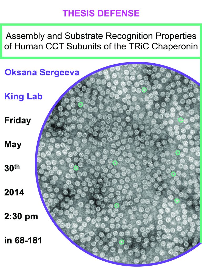
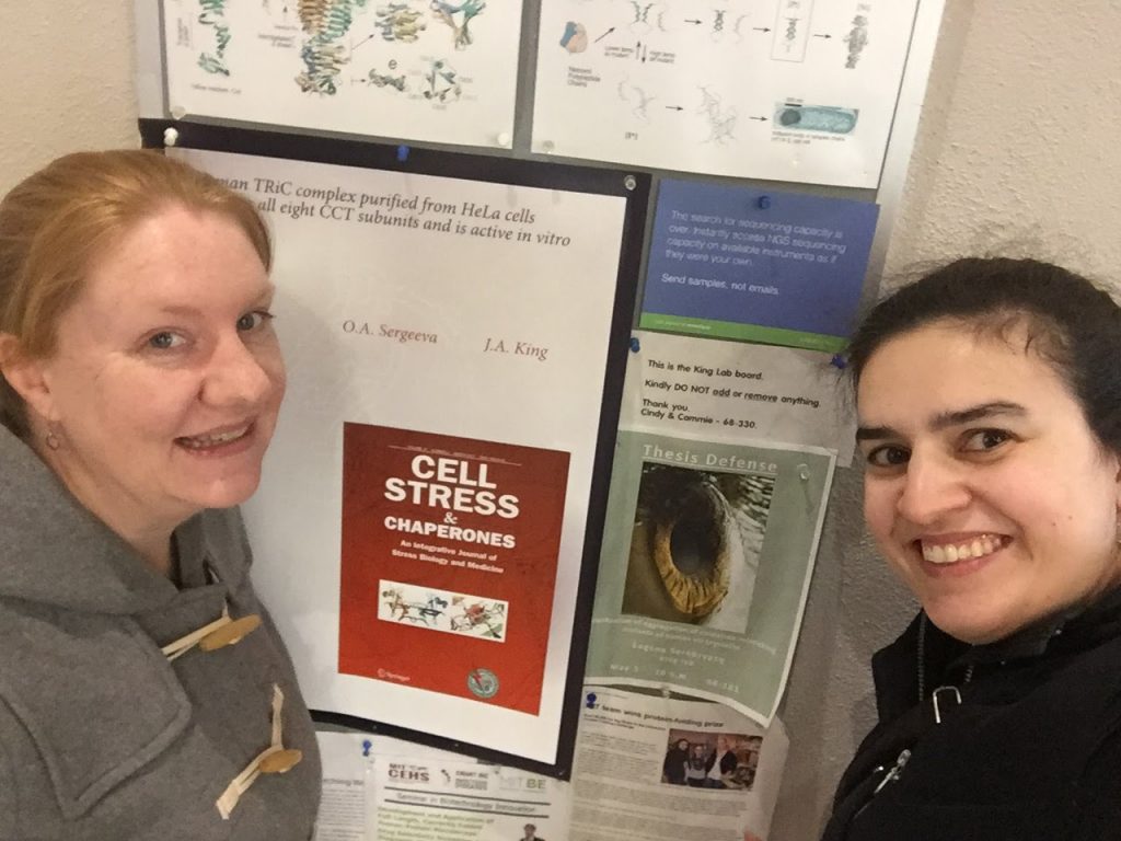
I started off working on crystallins and the archaeal chaperonin Mm-Cpn, a project started by Kelly Knee1. We shifted to the human chaperonin called TRiC, first purifying out of human cells2, and then actually purifying the human chaperonin subunits out of E. coli3. The fact that we could get some TRiC subunits to make functional complexes in vitro made it possible to study a slew of other properties of this human chaperonin. This included substrates other than crystallins: actin and the huntingtin protein that was responsible for Huntington’s disease4. I have always been interested in disease mutations, so I was able to direct the project to study mutations of the chaperonin subunit that lead to neuropathies5.6. One of my regrets is not being able to share more of this study at conferences as the results were so cool, but it was published right at the end of my time in Jon’s lab. In my last year in the lab, we finished one last study with Cammie Haase-Pettingell in which we looked at how the human chaperonin may assemble from its subunits, something that I didn’t write-up and submit until way later7.

One of the highlights of being in Jon’s lab was how supportive he was in everything, especially career progression. Working on chaperonins allowed me to be part of the Nanomedicine Consortium which included yearly conferences and opportunities to collaborate with other labs working on CryoEM, x-ray crystallography, and cell biology. I travelled to our center conferences, often taking place in San Diego, and also had stints in labs in Houston, Berkeley, and Irvine learning new techniques. In return, some of those scientists also came to Cambridge to learn our techniques. These interactions helped me better communicate and extend my network past our lab. In addition, he also encouraged me to attend various Gordon and FASEB conferences and a conference in Mexico, giving me a great perspective of my work in the larger biochemistry/protein folding field. Jon was also very big on mentoring undergraduates in the lab, allowing me the opportunity to take on students, which I found out I really enjoyed. Meme Tran was an undergraduate student who worked on the neuropathy project and really prospered in the lab, as a supplement to her studies.
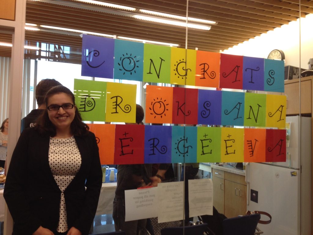
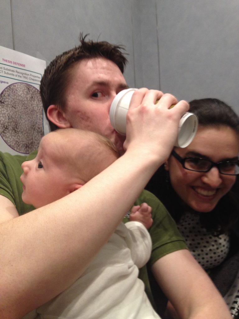
Jon was also very supportive of our lives outside the lab and science. Not only did I get married during my time in Jon’s lab, but I also had a baby. Chase was almost 7 weeks old when I defended my thesis at the end of May 2014. Jon knew that my husband had to go to Switzerland to finish his PhD, so he was very good about introducing me to European professor colleagues of his at conferences and asking them about cell biology professors who would make good postdoc mentors. Jon actually sent letters of recommendation directly to each professor a week before I sent them my application, a strategy that I hadn’t heard of but was very effective. In fact, the professor’s lab that I joined for my postdoc mentioned multiple times how much of an impact that letter made on her and how caring of a mentor he must have been for me during my PhD.
After graduating from Jon’s lab, I moved to Lausanne, Switzerland for a postdoc in cell biology. There, I studied host pathways involved in pathogen entry, extending into the fields of exosomes, post-translational modifications8, rare disease9, and during the pandemic, SARS-CoV-210. After a lengthy post-doc that got extended by the pandemic, I finally made my way back to the US in 2021 to start a job in biotech in Connecticut studying immuno-oncology. Moving so easily (and confidently) from biochemistry to cell biology to immunology is really a testament to how well the education I received in Jon’s lab prepared me for being a scientist. Jon gave me the foundation and skills to be able to go do any type of science to the level of rigor and deliberation he instilled in me, and for that I will be ever thankful.
- Sergeeva OA, Yang J, King JA, Knee KM. Group II archaeal chaperonin recognition of partially folded human γD-crystallin mutants. Protein Sci. 2014 Jun;23(6):693-702. doi: 10.1002/pro.2452.
- Knee KM, Sergeeva OA, King JA. Human TRiC complex purified from HeLa cells contains all eight CCT subunits and is active in vitro. Cell Stress Chaperones. 2013 Mar;18(2):137-44. doi: 10.1007/s12192-012-0357-z.
- Sergeeva OA, Chen B, Haase-Pettingell C, Ludtke SJ, Chiu W, King JA. Human CCT4 and CCT5 chaperonin subunits expressed in Escherichia coli form biologically active homo-oligomers. J Biol Chem. 2013 Jun 14;288(24):17734-44. doi: 10.1074/jbc.M112.443929.
- Darrow MC, Sergeeva OA, Isas JM, Galaz-Montoya JG, King JA, Langen R, Schmid MF, Chiu W. Structural Mechanisms of Mutant Huntingtin Aggregation Suppression by the Synthetic Chaperonin-like CCT5 Complex Explained by Cryoelectron Tomography. J Biol Chem. 2015 Jul 10;290(28):17451-61. doi: 10.1074/jbc.M115.655373.
- Sergeeva OA, Tran MT, Haase-Pettingell C, King JA. Biochemical characterization of mutants in chaperonin proteins CCT4 and CCT5 associated with hereditary sensory neuropathy. J Biol Chem. 2014 Oct 3;289(40):27470-80. doi: 10.1074/jbc.M114.576033.
- Pereira JH, McAndrew RP, Sergeeva OA, Ralston CY, King JA, Adams PD. Structure of the human TRiC/CCT Subunit 5 associated with hereditary sensory neuropathy. Sci Rep. 2017 Jun 16;7(1):3673. doi: 10.1038/s41598-017-03825-3.
- Sergeeva OA, Haase-Pettingell C, King JA. Co-expression of CCT subunits hints at TRiC assembly. Cell Stress Chaperones. 2019 Nov;24(6):1055-1065. doi: 10.1007/s12192-019-01028-5.
- Sergeeva OA, van der Goot FG. Anthrax toxin requires ZDHHC5-mediated palmitoylation of its surface-processing host enzymes. Proc Natl Acad Sci U S A. 2019 Jan 22;116(4):1279-1288. doi: 10.1073/pnas.1812588116.
- Sergeeva OA, van der Goot FG. Converging physiological roles of the anthrax toxin receptors. 2019;8. doi: 10.12688/f1000research.19423.1.
- Mesquita FS, Abrami L, Sergeeva O, Turelli P, Qing E, Kunz B, Raclot C, Paz Montoya J, Abriata LA, Gallagher T, Dal Peraro M, Trono D, D’Angelo G, van der Goot FG. S-acylation controls SARS-CoV-2 membrane lipid organization and enhances infectivity. Dev Cell. 2021 Oct 1;. doi: 10.1016/j.devcel.2021.09.016.